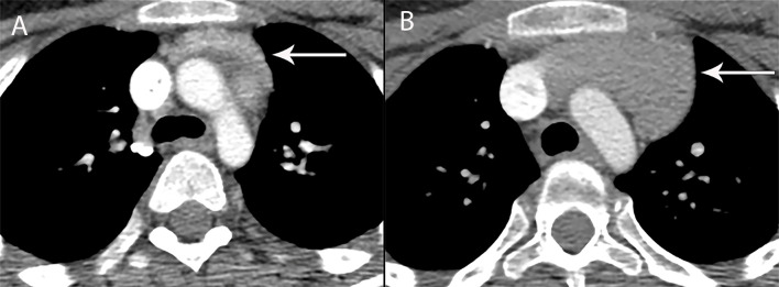
Figure 6.
13 year old boy with chondroblastic osteosarcoma of the femur, treated with methotrexate, doxorubicin, cisplatin (A) CT shows the normal thymus (arrow) at baseline. (B) CT 4 months later shows increase in size of the thymus consistent with rebound hyperplasia (arrow). Enlargement of the thymus gland due to hyperplasia during the recovery phase from physical stress such as after chemotherapy or recovering from burns, does not displace or change the contour of vessels surrounding it. In the appropriate clinical context of thymic hyperplasia, CT is adequate for diagnosis and MRI is not needed for confirmation.

