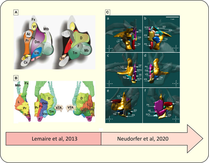
Figure 4.
Representative diagram of the advances in the parcellation of the hypothalamus. (A) Schematic drawing of hypothalamic nuclei, lateral view from the midline. Left, nuclei directly located under the wall of the third ventricle; right, more deeply located nuclei up to the lateral region (52). (B) 3D overview of hypothalamic nuclei, constructed from a 3D high-field MRI data set. Left, frontal view; middle, lateral view; right, medial view. VTA, ventral tegmental area; Ac, anterior commissure; D, dorsal or posterior nucleus: Dm, dorsomedial nucleus; Fx, fornix; F, fornix nuclei; If, infundibular (arcuate) nucleus; L, lateral nucleus; Mb, mammillary body; Mtb, Fmillo-thalamic bundle; Cx, optic chiasma; Pv, paraventricular nucleus; Pr, preoptic nuclei; Sc, suprachiasmatic (ovoid) nucleus; Vf, ventricular foramen; So, supraoptic (tangential) nucleus; Tm, tuberomamillaris (mamilloinfundibularis) nucleus; Vm, ventromedial (tuber principal) nucleus (52). (C) 3D reconstruction of hypothalamic nuclei and their neuroanatomical relationships. (a) top view, (b) bottom view, (c) frontal view, (d) occipital view, (e) sagittal view depicting the outer surface of hypothalamic nuclei, (f) sagittal view depicting the inner surface of hypothalamic nuclei. AH, anterior hypothalamic area; AN, arcuate nucleus; DP, dorsal periventricular nucleus; DM, dorsomedial hypothalamic nucleus; LH, lateral hypothalamus; MPO, medial preoptic nucleus; PA, paraventricular nucleus; PE, periventricular nucleus; PH, posterior hypothalamus; SCh, suprachiasmatic nucleus; SO, supraoptic nucleus; TM, tuberomammillary nucleus; VM, ventromedial nucleus (4).

