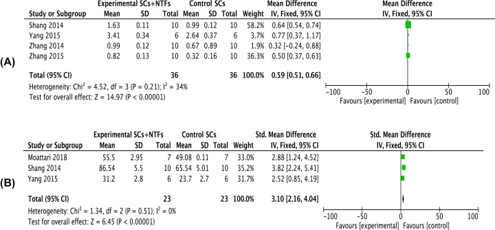
Figure 5. Forest plots of nerve transmission electron microscopy indicators.
(A) Showed the meta-analysis of thickness of myelin sheath of the affected limb after stem cell treatment and stem cell combined neurotrophic factor treatment in rats with sciatic nerve injury at 8 weeks. (B) Showed the meta-analysis of number of myelinated nerve fibers of the affected limb after stem cell treatment and stem cell combined neurotrophic factor treatment in rats with sciatic nerve injury at 8 weeks. Experimental group: stem cells combined with neurotrophic factors treatment group; Control group: stem cells treatment group. Neurotrophic factor combined with stem cell in the treatment of nerve transmission electron microscopy indicators were significantly better than that in the stem cell treatment group, the difference was significant (P<0.05).

