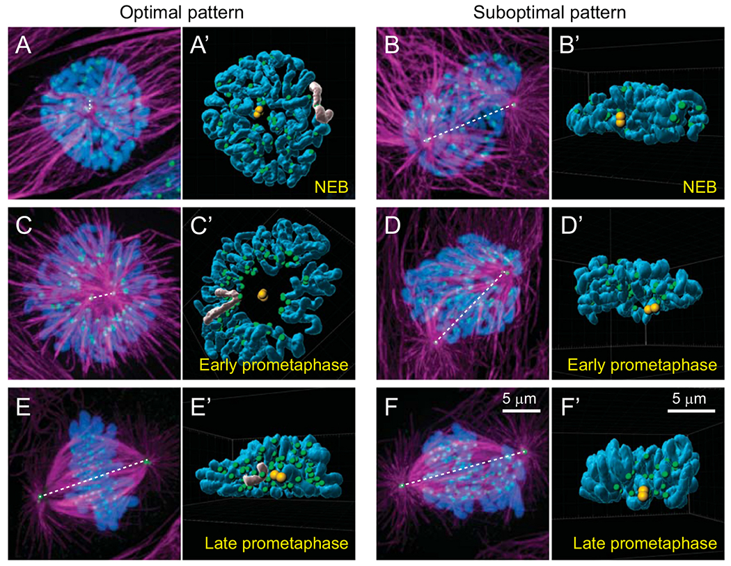
Fig. 2.

Steric constraints arising from different relative positions of centrosomes and chromosomes at the onset of spindle assembly. (A,B) The number of kinetochores with a direct line of sight to the spindle poles depends on whether the centrosomes separate along the shorter vs. longer axis of the nucleus. In the ‘Optimal pattern’ (A), chromosomes form a thin layer orthogonal to the spindle axis while in the ‘Suboptimal pattern’ (B) many kinetochores are shielded from microtubules by chromosome arms (compare the number of green spots visible in A’ vs. B’). (C,D) Optimal positioning of centrosomes at NEB promotes toroidal distribution of chromosomes during early prometaphase (C). Within the toroid, kinetochores (green) reside near the surface of a microtubule-dense ‘nascent spindle’ (magenta) while chromosome arms are ejected outwards (compare shape of chromosomes greyed in A’ vs. C’). Suboptimal pattern of centrosome orientation leads to multiple layers of chromosomes. (E,F) In the Optimal pattern, chromosomes rapidly repopulate the inner parts of the spindle (E,E’). In contrast, wider distribution of chromosomes in the Suboptimal pattern delays chromosome alignment at the spindle equator (F,F’). (A-F) Maximal intensity projections of the entire cell volumes with microtubules shown in magenta (α-Tubulin), kinetochores and centrosomes in green (CenpA-GFP and Centrin1-GFP), and chromosomes in blue (Hoechst 33342). Orientation of the spindle axis is denoted by a dashed white line. (A’-F’) Surface-rendered models of presenting positions of chromosome arms (blue), kinetochores (green), and centrosomes (yellow) in 3D space. Each model is oriented to present the view from one centrosome towards the other.
