
Fig. 4.
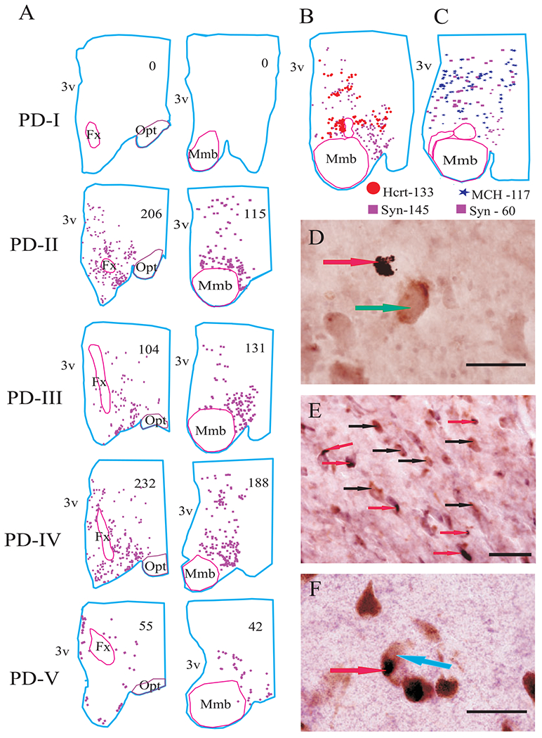
Distribution of alpha synuclein in the hypothalamus in different stages of PD. (A) Neurolucida mapping of alpha synuclein in PD stages with single immunostaining. (B) Mapping of Hcrt and alpha synuclein in double-labelled section. (C) Mapping of MCH and alpha synuclein in double-labelled section. Alpha synuclein was not colocalized with Hcrt and MCH cells (D and E), but it was colocalized with neuromelanin pigmented cells in substantia nigra (F). Arrows: red—alpha synuclein, green—Hcrt cell, black—MCH cells and blue—neuromelanin pigmented cell. Scale bars—50 μm.