
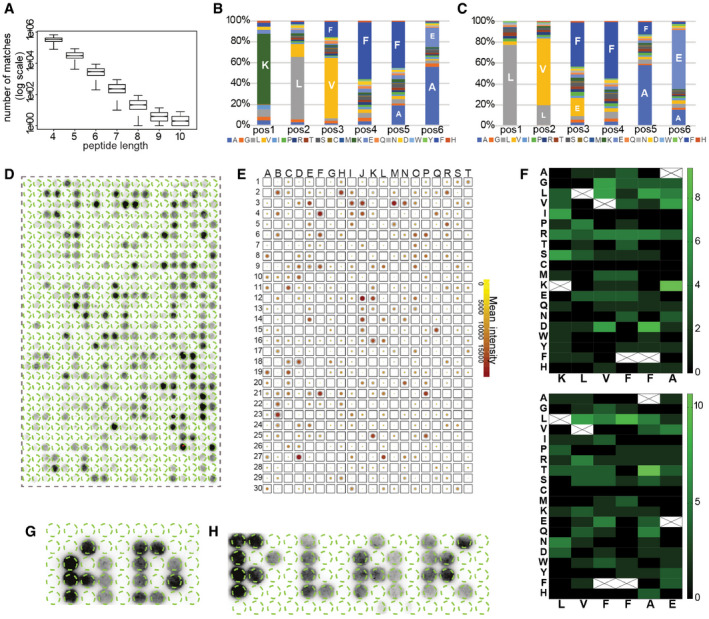
Figure 2. Aβ binding to APR homologues derived from human proteins.
-
ASequence similarity in combination with peptide length for 1,000 random proteins derived from human proteome. Graph: bottom and top of the boxes are the first and third quartiles, central band represents median, whiskers encompass minimum and maximum.
-
B, CDistribution of amino acids in homologues to Aβ KLVFFA (B) and LVFFAE (C) proteins.
-
DBinding of Biot‐Aβ1‐42 to homologue peptides derived from ˜ 520 randomly selected proteins.
-
ESummary of Biot‐Aβ1‐42 binding throughout eight membranes, colour indicates the mean between membranes and the size of the outline of the standard deviation.
-
FHeatmap of amino acid substitutions in membrane hits.
-
G, HMembrane top binders spell AD and PLAK, white space consists of random sequences. (sequences in Appendix Table S2).