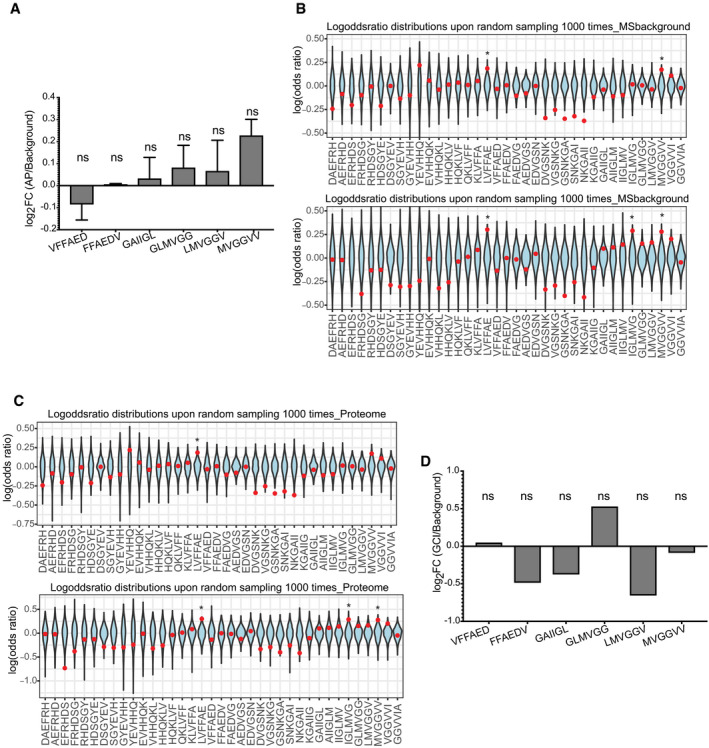
Figure 5. No over‐representation is observed in amyloid plaques from APP/PS1 mouse or in Glial cytoplasmic inclusions from.

- Over‐representation of Aβ APRs in amyloid plaques of APP/PS1 mouse brains (mean ± SD) from two biological replicates. (statistics: hypergeometric test with Bonferroni correction).
- Log‐odd ratio of random sampling from mass spectrometry background for both replicates. Red dot indicates the true values of analysis. (statistics: Z‐test) *P ≤ 0.05.
- Log‐odd ratio of random sampling from mouse proteome for both replicates. Red dot indicates the true values of analysis. (statistics: Z‐test) *P ≤ 0.05.
- No over‐representation of Aβ APRs was observed in proteins from Glial cytoplasmic inclusions (α‐synuclein aggregates). (statistics: hypergeometric test with Bonferroni correction).
