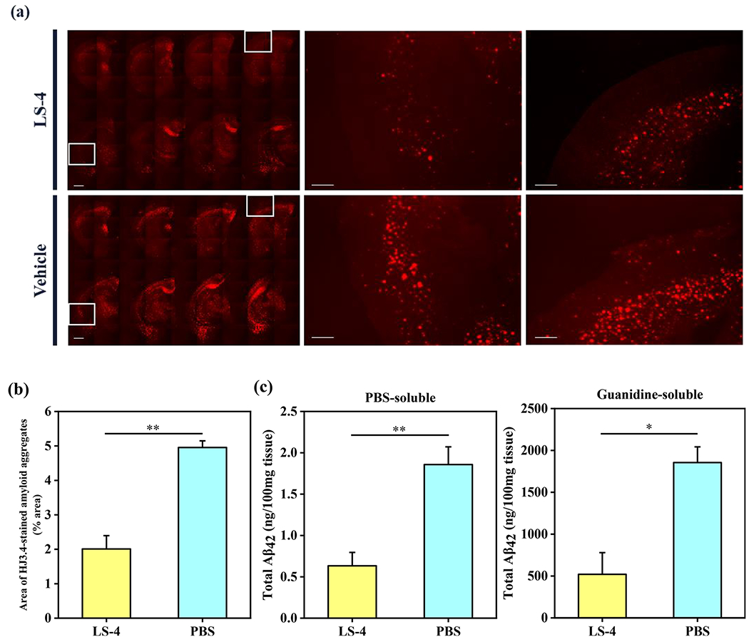
Figure 9.

Reduction of cerebral amyloid pathology by LS-4 in 5xFAD mice. The brain tissues were collected from the 3-month old 5xFAD mice after 30 days i.p. injections of LS-4 or vehicle. (a) Representative fluorescence microscopy images of the CF594-HJ3.4 antibody-stained brain sections from 5xFAD mice treated with LS-4 and vehicle. Scale bar = 500 μm. (b) Total area of HJ3.4-staining amyloid plaques in the brain sections from 5xFAD mouse treated with LS-4 and vehicle. The area of antibody-stained amyloid plaques was selected from 8 brain sections per mice. For each brain section, five random areas across the cortex regions were chosen. Error bars represent the standard deviation (LS-4-treated mice, n = 4, vehicle-treated mice, n = 3), and the statistical analysis was evaluated according to one-way ANOVA (** p < 0.01). (c) The bars indicate the amount of PBS-soluble (left) and guanidine-soluble (right) Aβ42 peptide levels from brain tissues. Error bars represent standard deviations (LS-4 treated mice, n = 5, vehicle-treated mice, n = 3), and the statistical analysis was evaluated according to one-way ANOVA (** p < 0.01, * p < 0.05).
