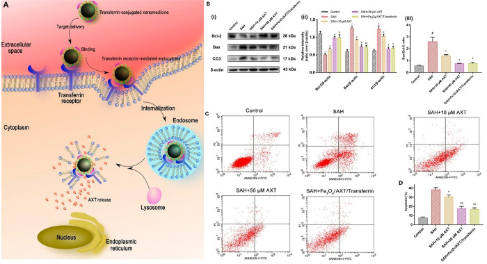
Figure 8.
(A) Schematic of the entry mechanism of transferrin-modified and -nonmodified nanoparticles to neurons through receptor-mediation followed by the degradation of nanoparticles and AXT release. (B) Assessment of neural damage after exposure to oxyhemoglobin for pure AXT and the transferrin-modified AXT-loaded nanoparticles as follows: (i) Western blots, (ii) relative intensity analysis of Bax/ß-actin, Bcl-2/ß-actin, and cleaved caspase-3 (CC3)/ß-actin, and (iii) Bax/Bcl-2 ratio for different samples. (C) Cell apoptosis results after oxyhemoglobin exposure. (D) Apoptotic ratio of cells related to each group. #p < 0.05 vs control group; p < 0.05 vs subarachnoid hemorrhage (SAH) group; p < 0.01 vs SAH group. Reprinted from ref (131) with permission from Frontiers.

