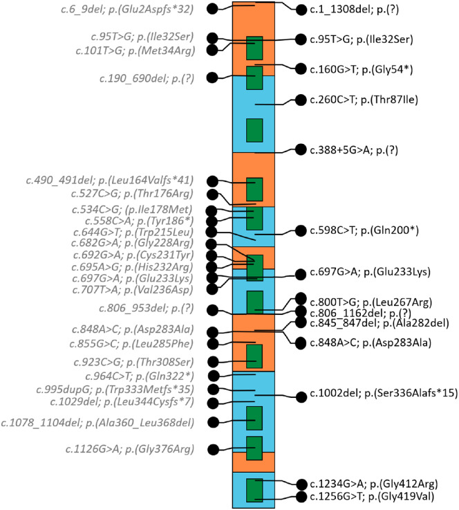
Figure 1.
Schematic representation of the SLC38A8 gene. The location of the variants found in this study patients is represented on the right and of those retrieved from literature on the left, in grey. Exons are illustrated in orange and blue, transmembrane domains in green. Deletions are depicted at the location of the first deletion coordinate.

