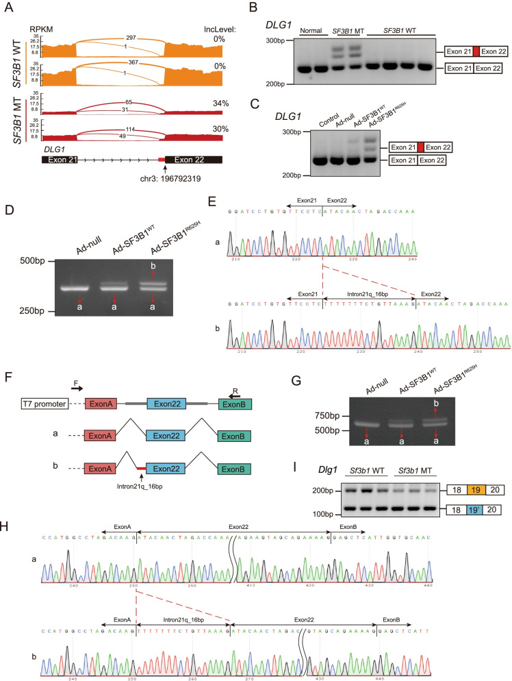
Fig. 3.
Mutant SF3B1 caused aberrant splicing of DLG1. A Sashimi plots of alternative 3′ splice sites of DLG1 in representative human prolactinoma with or without SF3B1 mutations. B RT-PCR of DLG1 splicing events in human prolactinoma samples with or without SF3B1 mutation. The 3′ splice sites used are shown in the schematic. C RT-PCR results of aberrant DLG1 splicing in primary human prolactinoma cells infected with adenovirus carrying SF3B1-WT and SF3B1-R625H vectors. D RT-PCR of DLG1 splicing in 293 T cells infected with adenovirus carrying SF3B1-WT and SF3B1-R625H vectors. Band a: canonical DLG1 transcript; Band b: aberrant DLG1 transcript. E The sequences of gel-purified fragments showing the canonical DLG1 transcript (a) and aberrant transcript (b). F Schematic diagram of the minigene construct and schematic diagram of Sanger sequencing of RT-PCR products. G RT-PCR results of DLG1 splicing in 293 T cells co-transduced with the minigene reporter and adenovirus carrying SF3B1-WT and SF3B1-R625H vectors. H The sequence of gel-purified fragments. The canonical DLG1 transcript (a) and aberrant DLG1 transcript (b) were observed in the SF3B1-WT and SF3B1-R625 groups, respectively. I RT-PCR results of Dlg1 splicing in Sf3b1 mutant GH3 cells. The mutually exclusive splicing type is shown in the schematic

