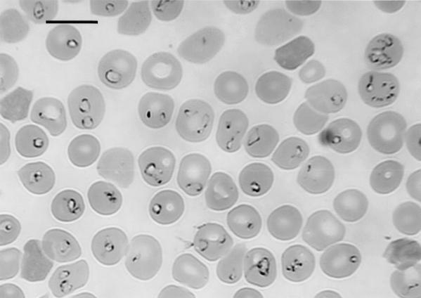
FIG. 3.
Photomicrograph of intraerythrocytic piroplasms in a hu-RBC-SCID mouse whose results are presented in Fig. 2. The Giemsa-stained thin-smear blood film was prepared 19 days after inoculation of the patient's blood specimen, when human RBCs made up 87% of the peripheral RBCs of the mouse. Bar, 10 μm.

