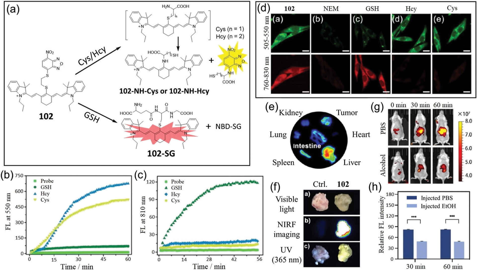
Fig. 18.

(a) Response mechanism of 102 for discriminating Cys/Hcy and GSH with two distinct emission patterns. (b and c) Time-dependent emission changes at 550 nm (b) and 810 nm (c) of 102 with the addition of GSH, Hcy and Cys, respectively. (d) Fluorescence images of U87 cells incubated with 102 (da); with N-ethylmaleimide (NEM) and then 102 (db); with NEM and then with GSH (dc), Hcy (dd), or Cys (de), followed by 102. (e) Ex vivo fluorescence images of isolated organs from the tumor-bearing mice at 8 h post-injection of 102. (f) Photos of tissue portions of tumors stained by PBS as control or 102, respectively, under visible light, or fluorescence at 760 nm or 365 nm excitation. (g) Representative NIR fluorescence images of mice (oral gavage 2 g kg−1 alcohol) were collected at 0, 30, and 60 min post-injection of 102. (h) Semi-quantitative analysis of the NIR fluorescence intensity of mice in (g). (b)–(h) are adapted with permission from ref. 184. Copyright 2020 Royal Society of Chemistry.
