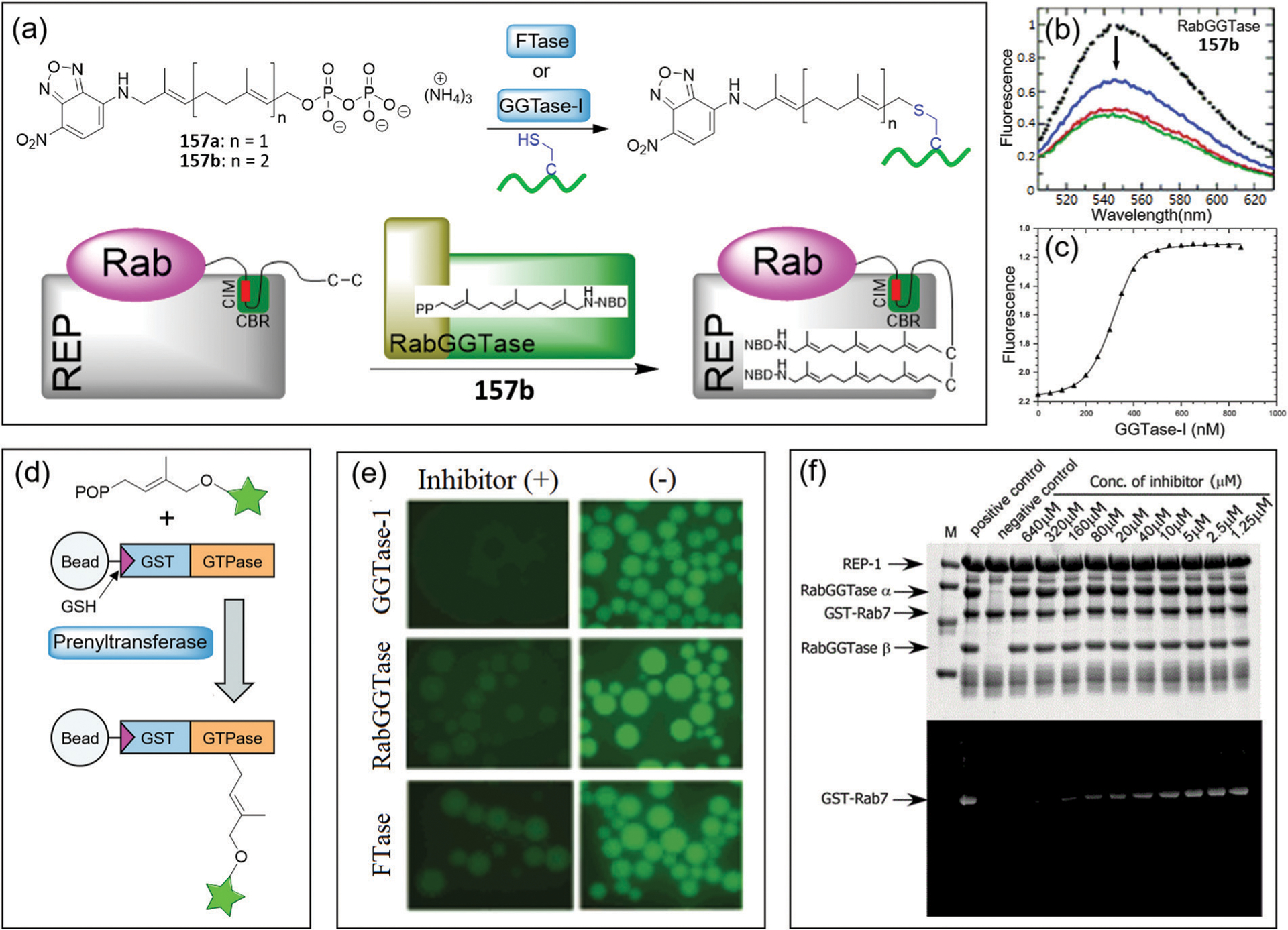
Fig. 50.

(a) Illustration of the prenylation reactions based on probes 157a–157b. (b) Emission spectra of 157b in the absence (black filled circles) and presence of RabGGTase (color lines). (c) Competitive titration of the 157b and GGPP mixture with increasing concentrations of GGTase-I. (d) Principle of the on-bead microscopic assay for identification of prenyltransferse inhibitors. (e) Fluorescence of GST-GTPase loaded beads after prenylation with probes 157a–157b in the presence or absence of putative inhibitors. (f) SDS-PAGE for the prenylation reactions containing various concentrations of inhibitor, or without RabGGTase as a negative control. The top and bottom panels for the gel after Coomassie blue staining and the fluorescent scan gel, respectively. (b)–(f) are adapted with permission from ref. 279. Copyright 2006 American Chemical Society.
