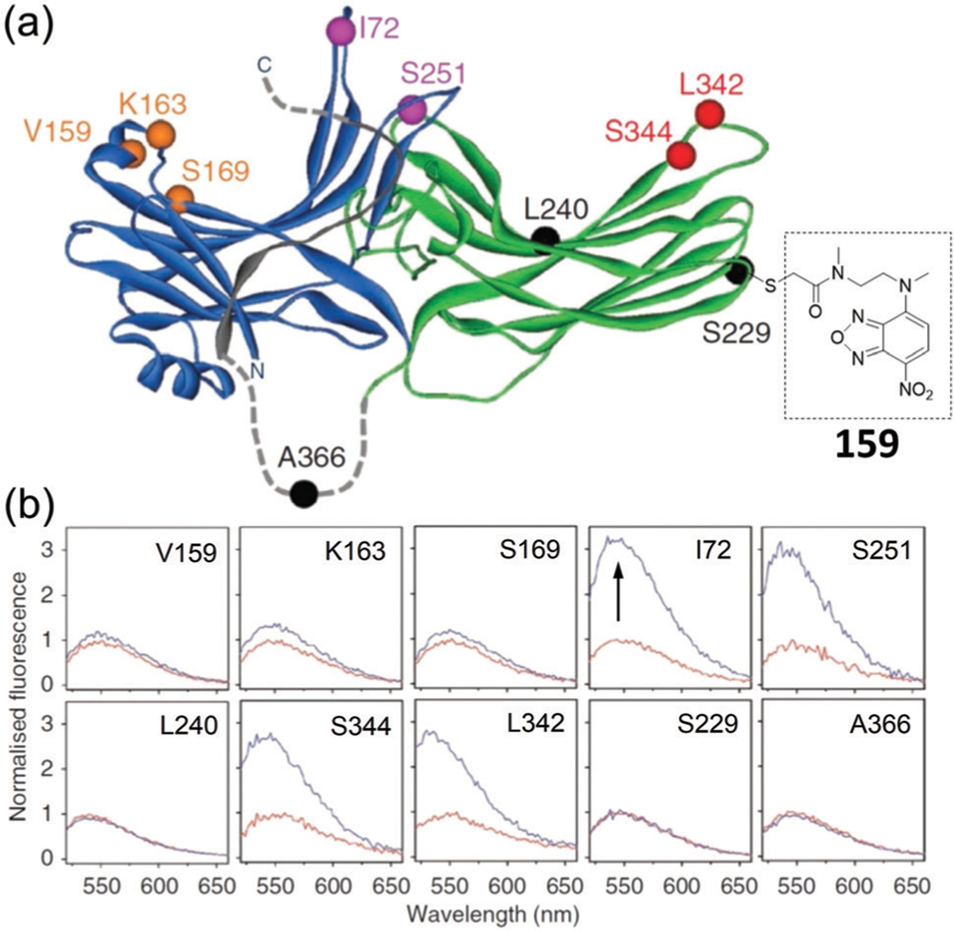
Fig. 53.

(a) Location of single Cys substitutions and chemical structure of the attached NBD fluorophore on a model of arrestin. (b) For each panel, NBD-labelled arrestin was mixed with a four-fold excess of dark-state rhodopsin. Adapted with permission from ref. 296b. Copyright 2012 Nature Publishing Group.
