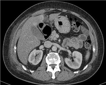
Figure 1 .

Contrast-enhanced CT scan shows a 32 × 22-mm oval-shaped fat density lesion with peripheral hyperattenuation and central hyperdense dot adjacent to falciform ligament. There is surrounding oedema and inflammation. Gall bladder is noted to be unremarkable. Features represent intraperitoneal focal fat infarction of fatty appendage of falciform ligament.
