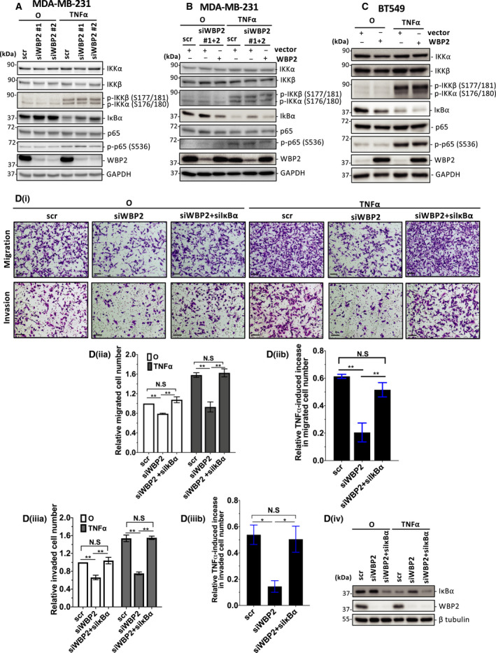
Fig. 5.

WBP2 limits IκBα protein level to drive TNF‐α‐induced migration and invasion. (A–C) Western blot analysis of NF‐κB signaling components upon 15min of TNF‐α stimulation (A) WBP2 was knocked down in MDA‐MB‐231 cells using two different siRNAs (siWBP2#1 and siWBP2#2) or scrambled (scr) siRNA control. (B) MDA‐MB‐231 cells were cotransfected with either scrambled (scr) or pooled siWBP2 (siWBP2#1 + 2), along with vector or WBP2‐expressing plasmid. (C) BT549 cells were transfected with either WBP2‐expressing plasmid or vector control. For A‐C, GAPDH was probed as a loading control. (D) Diminished cell migration and invasion caused by loss of WBP2 expression were reverted by simultaneous loss of inhibitor of nuclear factor of kappa beta alpha (IκBα). (i) Representative images of migrated and invaded MDA‐MB‐231 cells. Cells from 10 random fields of each sample were imaged. Magnification: 10×, scale bar (black): 250 µm. (ii) (a) Relative number of migrated cells were quantified and normalized to unstimulated scr control. O annotate cells unstimulated with TNF‐α. (ii) (b) Fold increase in migrated cells induced by TNF‐α. (iii) (a) Relative invaded cell number was quantified and normalized to unstimulated scr control (iii) (b) Fold increase in invaded cells induced by TNF‐α. Data are represented as mean ± SEM, n = 3. *P < 0.05; **P < 0.01 (one‐way ANOVA followed by post hoc Bonferroni test). (iv) WBP2 and IκBα knockdown in was validated in Western blot. β‐Tubulin was used as a loading control.
