
Figure 1.
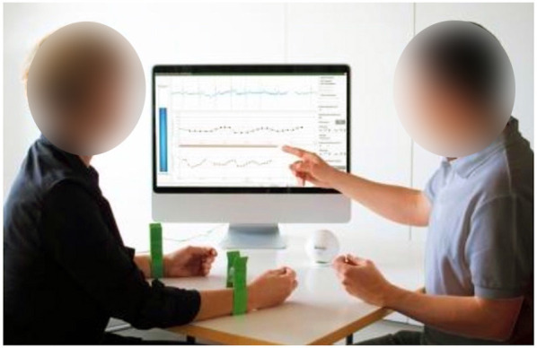
Evaluation of HRV with Deep Breathing Test by HRV Scanner. The patient, who is wearing wrist ECG electrodes, and the investigator are facing the computer screen; a bar instructing the patient on how to breathe is placed on the left side of the measurement window. The incoming biosignals, as well as the progression chart and measurement overview, are displayed in the center; HRV parameters are set in the configuration window on the right (representative picture–source: Mega HRV-Scanner Flier 02__2013 E DB-v.1.1).