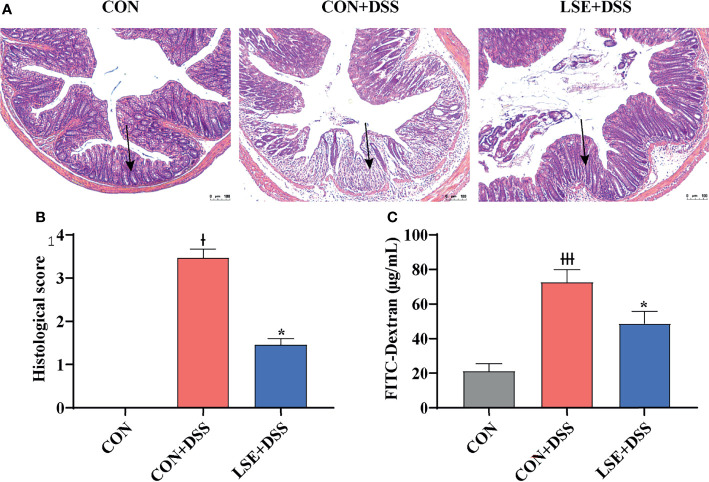
Figure 2.
Effects of LSE supplementation on histopathological changes in DSS-induced mouse colon. (A) H&E staining images of each group. (B) Histopathological score of each group. (C) Serum FITC-dextran levels of each group. Data are presented as mean ± SEM, n = 4. * P < 0.05 vs. the CON+DSS group, †P < 0.05 vs. the CON group, and †††P < 0.001 vs. the CON group.

