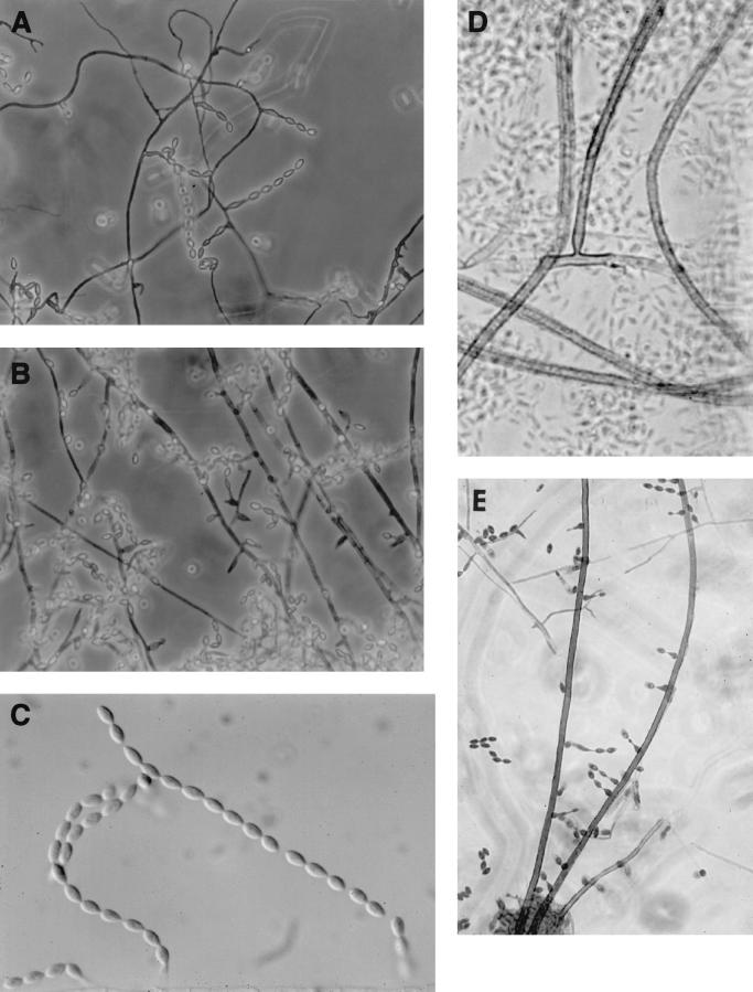
FIG. 5.
Microscopic morphology. (A) Basally inflated phialides of case isolate, UTHSC 99-2508, occurring along the sides of thin-walled, hyaline-to-pale brown septate hyphae (magnification, ×306). (B) Proliferating phialides of the case isolate, UTHSC 99-2508 (magnification, ×306). (C) Long chains of limoniform-to-fusiform, one-celled, smooth, hyaline conidia of the case isolate, UTHSC 99-2508 (magnification, ×613). (D) Echinulate, brown, prostrate conidiophore of isolate UTHSC 96-2378, anchored by a definite basal hyphal cell (magnification, ×580). (E) Basally inflated phialides of dog isolate R-3122, borne along the sides of brown, echinulate conidiophores (magnification, ×306).

