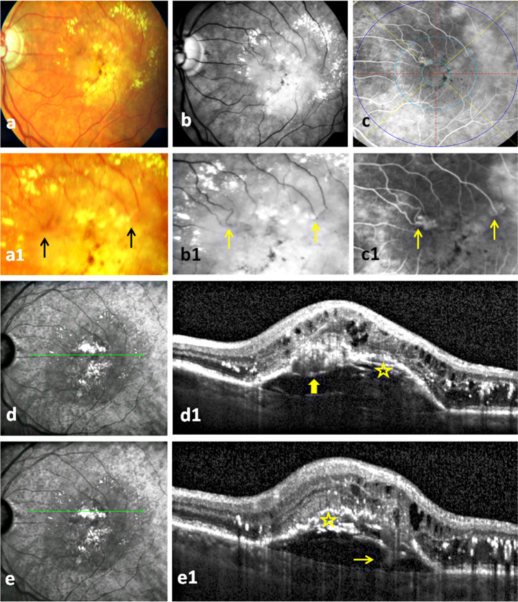
Fig. 1.
Bifocal macular neovascularization type 3 (MNV3): The first row presents the color fundus photo, red free, and fluorescein angiography (FA) images of massive hard exudates and central pigmentary clumping in a patient with bifocal MNV3 (a–c). Two MNV3 lesions with focal hyperfluorescence are located in the inner field of ETDRS (i.e., between 500 and 1500 µm from the fovea) in the temporal and nasal parts of the upper half of the macula on an early phase FA (c). The second row illustrates bigger images of the first row images which demonstrate the distinguishing retinal arteriolar-venular anastomosis at the nasal lesion and abrupt turning down of an arteriole at the temporal one (2 arrows) (a1–c1). Of course, FA is superior to other two modalities in identifying these vascular changes (c1). The near infrared images and optical coherence tomography scans at (1) the location of the nasal lesion present the characteristic interrupted PED with an overlying hyperreflective mass (thick arrow), intraretinal cysts, and extensive hard exudates (d, d1). Note the wide spread of pseudodrusen in the outer macula in near infrared images which is very common to find in MNV3 (d). (2) At the location of the temporal lesion, they demonstrate the same OCT changes of the nasal lesion and the extension of the hyperreflective band from the retina through the PED to the choroid referring to the retinal-choroidal anastomosis (thin arrow) (e1). The onion sign (asterisk) underneath the PED in both scans refers to the extensive lipid deposits (d1, e1)

