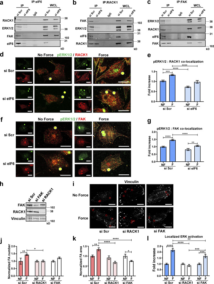
Figure 6.
eIF6 stabilizes mechanocomplexes. ECs were transfected with si Scr or si eIF6. (a) Immunoprecipitants, using an IgG- or eIF6-specific antibody, from si Scr and si eIF6 EC lysates were tested for association of RACK1, ERK1/2, and FAK; representative blots show eIF6-dependent associations (n = 3). (b) Immunoprecipitants, using an IgG- or RACK1-specific antibody, from si Scr and si eIF6 EC lysates were tested for association of ERK1/2, FAK, and eIF6; representative blots show eIF6-dependent associations (n = 3). (c) Immunoprecipitants, using an IgG- or FAK-specific antibody, from si Scr and si eIF6 EC lysates were tested for association of RACK1, ERK1/2, and eIF6; representative blots show eIF6-dependent associations (n = 3). (d–g) si Scr or si eIF6 ECs were exposed to force for 0 min (no force [NF]) or 30 min (force [F]). (d and f) Representative immunofluorescent micrographs showing colocalization of pERKT202/Y204 (green) and RACK1 (red; d), and pERKT202/Y204 (green) and FAK (red; f) following application of force. Magnetic beads are highlighted by white circles. Scale bars = 20 μm. (e and g) Image analysis quantification of colocalization of pERKT202/Y204 with RACK1 (e) and pERKT202/Y204 with FAK (g), using Pearson’s coefficient, following force (n > 30 cells across three separate experiments). (h) ECs were transfected with si Scr, si RACK1, or si FAK. Mechanical force was applied for 0 min (NF) or 30 min (F) to si Scr, si FAK, and si RACK1 ECs. (i) Representative superresolution immunofluorescent micrographs showing focal adhesions (vinculin; white) in ECs following force. Magnetic beads are highlighted by red circles. Scale bars = 20 μm. (j–l) Quantification of mean frequency of focal adhesions per cell (j), mean area of vinculin-positive focal adhesions (k), and localization of pERKT202/Y204 at focal adhesions (l; n > 30 cells across three separate experiments). Values in e, g, and j–l are mean ± SEM, and significance was determined by two-way ANOVA. *, P < 0.05; **, P < 0.01; ***, P < 0.001; ****, P < 0.0001. FA, focal adhesion; IP, immunoprecipitation; WCL, whole-cell lysate. Source data are available for this figure: SourceData F6.

