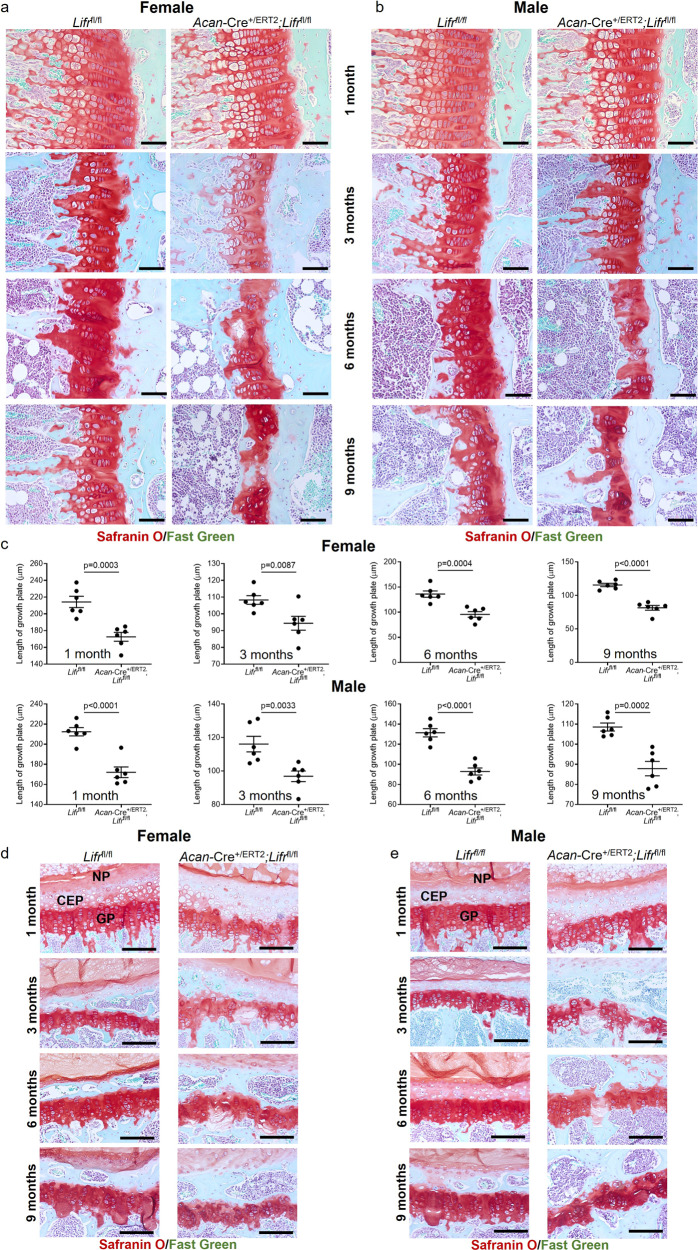
Fig. 8. Postnatal loss of Lifr in chondrocytes partially phenocopies Stat3 deletion.
Proximal tibial growth plates were significantly reduced in thickness at all ages examined following deletion of Lifr at P2/P3 in female (a, c) and male (b, c) mice; growth plate disruptions were not as severe as those observed in Stat3 and gp130 deletion mutants. Vertebral growth plates were mildly reduced in thickness, coupled with occasional disruption of integrity at later ages examined, in both female (d) and male (e) Acan-Cre+/ERT2;Lifrfl/fl mice as compared to Lifrfl/fl controls. AF annulus fibrosis, CEP cartilaginous endplate, NP nucleus pulposus. In all panels, scale bars = 50 µm; n = 6.

