Fig. 1.
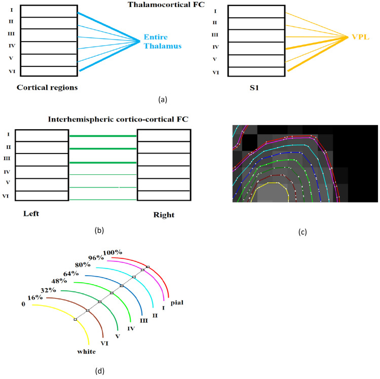
Illustration of our functional hypotheses that were motivated by previous invasive anatomical tract tracing studies. The width of the lines represent the strength of the connections. a Thamalocortical hypotheses: we hypothesized that FC between the entire thalamus and cortical layers I and VI will be significantly stronger than between the thalamus and other layers (blue, left panel). Furthermore, FC between somatosensory thalamus (ventral posterolateral nucleus, VPL) and layers IV, VI of the primary somatosensory cortex (S1) will be stronger than with other layers (yellow, right panel). b Cortico-cortical hypothesis: inter-hemispheric cortico-cortical FC between homologous regions in superficial layers (layers I–III) will be stronger compared to that in deeper layers (layers V–VI). c 6 surfaces plus white matter and pial surface overlayed on anatomical MRI (white matter surface: yellow, layer VI surface: brown, layer V: green, layer IV: lime, layer III: blue, layer II:, cyan, layer I: purple, and pial surface: red; the white dots are the vertices on these surfaces). d Illustration of the relative distance of 6 intermediate surfaces to white matter surface

