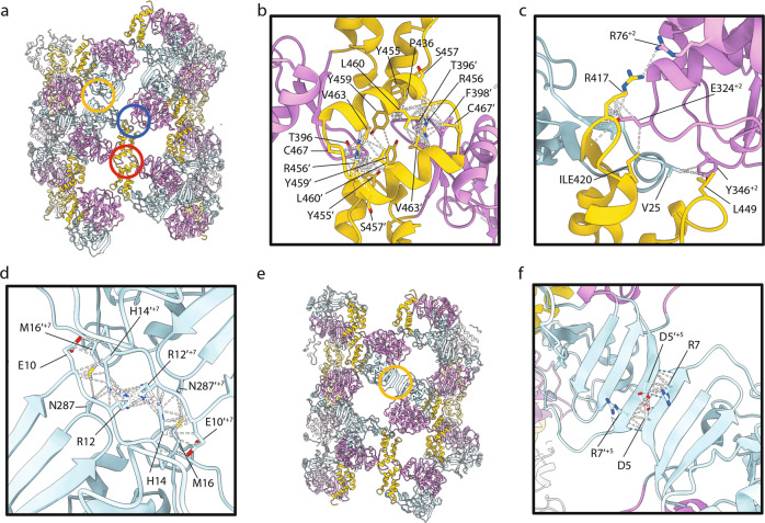
Fig. 6. Molecular interactions within the fibril.
a The pseudoatomic model for fibril N3 is shown as cartoons coloured according to the subdomain structure of 3Dpol; palm subdomain (plum), fingers subdomain (powder blue) and thumb subdomain (gold). Three interactions between monomers are highlighted: dimer interface (red circle), inter-dimer interface (blue circle) and inter-protofilament interface (yellow circle). Close-up views of the dimer interface b, inter-dimer interface c, and inter-protofilament interface d are presented for N3 with contacts/clashes highlighted by white dashed lines and sidechains of putative contact residues shown. e The pseudoatomic model of N9 is annotated to highlight the inter-protofilament interaction in this conformation (yellow circle), which is shown in close-up view with contacts/clashes highlighted with white dashed lines and putative hydrogen bonds indicated by blue dashed lines f.

