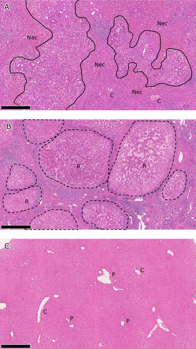
Fig. 1.
Pathological findings in A) ALF, B) LC, and B) HD livers. Representative findings are shown for each liver disease category. The scale bar represents 500 μm. A) Hepatocytes have been extensively destroyed to form bridging/panlobular necrotic areas (Nec), where collapsed hemorrhagic stroma, inflammatory cell infiltrates, and ductular reactions are seen. Native structures of the liver, including the portal venules (P) and central venules (C), are preserved. B) Liver structures are totally remodeled in the cirrhotic liver, which is characterized by nodules of regenerating hepatic parenchyma (R, regenerative nodules). Regenerative nodules are separated by dense fibrous septum with vascular vessels and inflammation. C) Intact portal venules (P) and central venules (C) are embedded in hepatic parenchyma in the normal liver. ALF, acute liver failure; LC, liver cirrhosis; HD, healthy donor.

