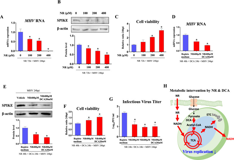
Fig. 6.
NR suppresses viral replication. Cells were infected by MHV (MOI 0.05) for 24 h following NR pre-treatment for indicated hours. Metabolite and chemicals were treated after serum free medium is changed to FBS containing medium. A, D RNA expression of MHV in cells treated with NR (indicated dose and time) and DCA (20 mM, 24 h) in replete medium (glucose 450 mg/dl, pyruvate 0.5 mM). Rplp0 was used for an internal control. B, E Western blot analysis and quantification of SPIKE in cells treated with NR (indicated dose and time) and DCA (20 mM, 24 h) in replete medium (glucose 450 mg/dl, pyruvate 0.5 mM). β-actin was used for an internal control. C, F Viability of cells incubated for 24 h with medium collected from MHV-infected cells which were treated as indicated. G Infectious virus titer (Log10PFU/ml) measured by plaque forming assay. H Illustration of metabolic intervention induced by NR and DCA. Values represent means ± SD. *p < 0.05. One-way ANOVA followed by a tukey’s multiple comparison test. All experiments were repeated at least 3 times

