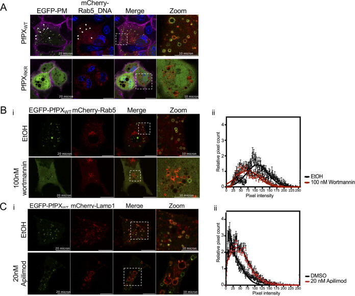
FIG 3.
The PX domain of PfPX1 likely binds selectively to PI3P. (A) Representative images of Cos-7 cells co-transfected with EGFP-PfPXWT or EGFP-PfPXRKR and mCherry-tagged early endosome marker Rab5. Blue: Hoechst-stained DNA. Magenta: plasma membrane stained with wheat-germ hemagglutinin-Alexa fluor 647. White arrows show colocalization of EGFP signal with mCherry signal indicating PfPXWT binding to early endosomes. Zoom panel corresponds to the insets showed in Merge panel. (B) Representative confocal images of Cos-7 transfected with EGFP-PfPXWT treated with ethanol (EtOH) or 100 nM Wortmannin. EGFP fluorescence is more cytosolic and reduced at mCherry-Rab5 positive structures in Wortmannin-treated cells (i). This reduction of EGFP signal is represented as histogram of intensity values (ii) indicating the reduction of pixel counts and intensity at mCherry-Rab5 positive structures upon Wortmannin treatment. (C) Representative confocal images of Cos-7 transfected with EGFP-PfPXWT treated with DMSO or 20 nM Apilimod (i). The EGFP-signal is represented as histogram of intensity values (ii) indicating a slight increase of pixel intensity at mCherry-LAMP-1 positive structures upon PIKfyve inhibition. The data in panels B and C are shown as the mean ± standard deviation with Gaussian fitting from three independent replicates, with a total of 40 to 60 vesicles.

