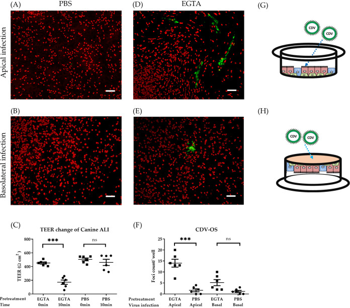
FIG 3.
Disruption of tight junctions enables CDV-OS infection of well-differentiated canine ALI cultures. Canine ALI cultures grown on filter supports with 0.4-μm pore size were inoculated by CDV-OS (MOI = 0.5). Prior to cell-free virion inoculation, cells were pretreated with PBS containing magnesium-calcium or with 100 mM EGTA in PBS (without magnesium-calcium) on the apical and basolateral sides. Before and after the incubation time of 10 min, the TEER values of the treated ALI cultures were measured (C). Following this pretreatment, CDV-OS was inoculated on the apical surface or onto the basolateral surface in an upside-down position for 2 h. On day 3 postinoculation, the ALI cultures were fixed by 3% PFA, and the ciliated cells were stained with antibodies against β-tubulin. The CDV-OS eGFP signals were visualized under the Nikon fluorescence microscope (A, B, D, and E), and the numbers of virus foci (green) were calculated and statistically analyzed (F). Schemes describing apical inoculation (G) and basolateral cell-free virion inoculation method with upside-down position (H). Bar, 100 μm. Statistical analysis: one-way ANOVA with Tukey’s post hoc test; ***, P < 0.001; ns, not significant.

