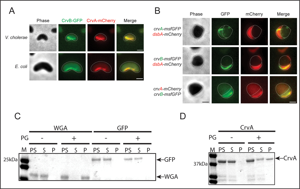
Figure 3. CrvA and CrvB break symmetry by forming a periplasmic filament that binds PG.
(A) Curved V. cholerae and E. coli cells expressing CrvB-GFP and CrvA-mCherry fusions. (B) Fluorescent CrvA and CrvB filaments with periplasmic DsbA-mCherry in V. cholerae after moenomycin treatment. (C,D) SDS-PAGE of PG containing (+) or PG lacking (−) pre-spin (PS), supernatant (S), and pellet (P) fractions from co-sedimentation assay with molecular weight standard (M) Representative of three independent experiments with similar results. (A,B) Dotted lines represent outline of cell body and periplasm. Scale bars are 1μm; images within each figure panel are to scale.

