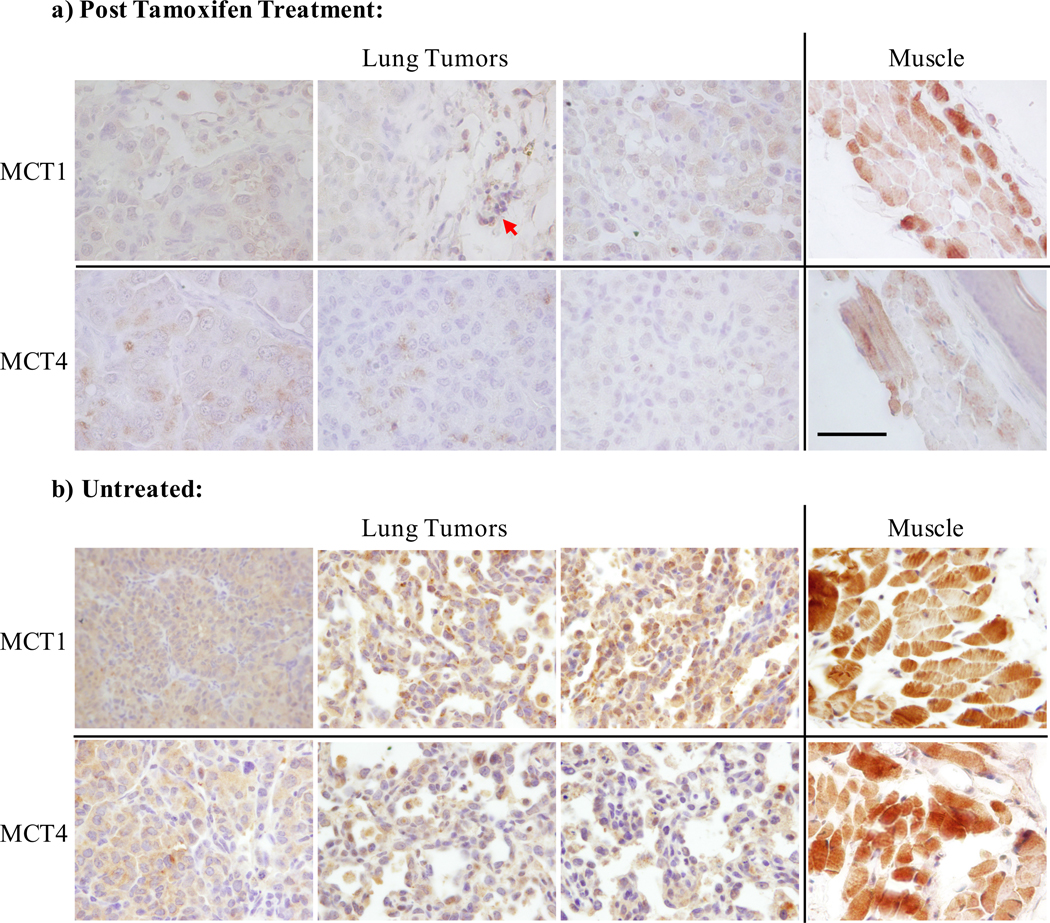
Figure 5.
IHC staining for the transporters MCT1 and MCT4 in three representative animals, a) following tamoxifen administration to induce deletion of LDH-A, and b) untreated animals. Tumors from the three mice shown in the upper panel are the same that contribute to data plot in Figure 4c. Robust staining of the musculature (rightmost panels) as well as mono-nuclear cells (red arrow) served as an internal control for the staining. Consistent with prior reporting, expression of the transporters was low. Slides were processed according to standard protocol for IHC with antibodies against MCT1 and MCT4. Scale bar = 100 μm.

