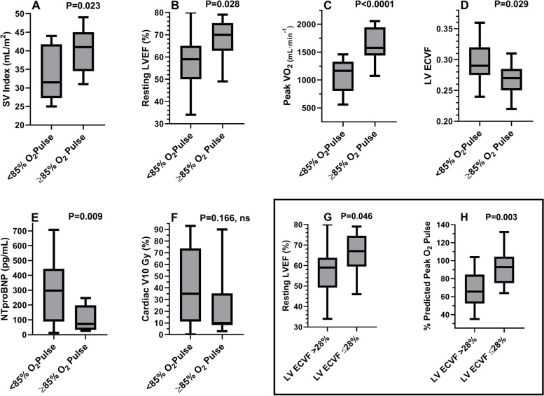
Fig. 3.
Differences between groups based upon percent-predicted peak exercise O2 pulse or median LV ECVF. Legend: Box and whisker plots (Panels A-F) demonstrating median (horizontal line within rectangular box), interquartile range, whiskers, and range between groups based upon %O2 pulse (< or ≥ 85%). Panel A: Lower SVI in those with peak O2 pulse < 85% of predicted. Panel B: Lower LVEF in those with a peak O2 pulse < 85% of predicted. Panel C: Lower peak VO2 in those with a peak O2 pulse < 85% of predicted. Panel D: Higher LV ECVF in those with a peak O2 pulse < 85% of predicted. Panel E: Higher NTproBNP levels in those with a peak O2 pulse < 85% of predicted. Panel F: Trend towards higher V10Gy in those with a peak O2 pulse < 85% of predicted. Panel G: Lower resting LVEF in those with LV ECVF values >median. Panel H: Lower peak %O2 pulse values in those with LV ECVF >median. Abbreviations: SVI = stroke volume index; O2 = oxygen; LVEF = left-ventricular ejection fraction; VO2 = oxygen consumption; ECVF = extracellular volume fraction; NTproBNP=N-terminal pro-brain natriuretic peptide; V10Gy = %volume of heart receiving ≥10 Gray

