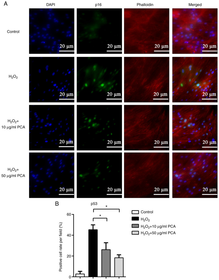
Figure 4.
PCA reduces p53 protein expression induced by H2O2 in NP cells. NP cells were treated with H2O2 and different concentrations of PCA and then cultured for 72 h. Subsequently, p53 protein expression was investigated by immunofluorescence. (A) Representative fluorescence microscopy images for the different groups (scale bar, 20 µm). (B) Bar graph indicating the p53-positive cell rate per field. Values are expressed as the mean ± standard deviation. *P<0.05. NP, nucleus pulposus; PCA, p-coumaric acid.

