Figure 5.
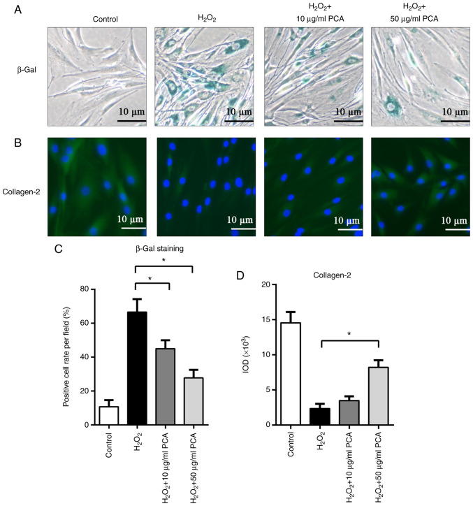
PCA inhibits senescence of NP cells induced by H2O2. NP cells were treated with H2O2 and different concentrations of PCA and then cultured for 72 h. (A) β-Gal staining of NP cells in the different groups. (B) Immunofluorescence staining for collagen-2 in the different groups (scale bars, 10 µm). Quantification of (C) β-Gal staining and (D) collagen-2 immunofluorescence staining. Values are expressed as the mean ± standard deviation. *P<0.05. NP, nucleus pulposus; PCA, p-coumaric acid; β-Gal, β-galactosidase; IOD, integrated optical density.

