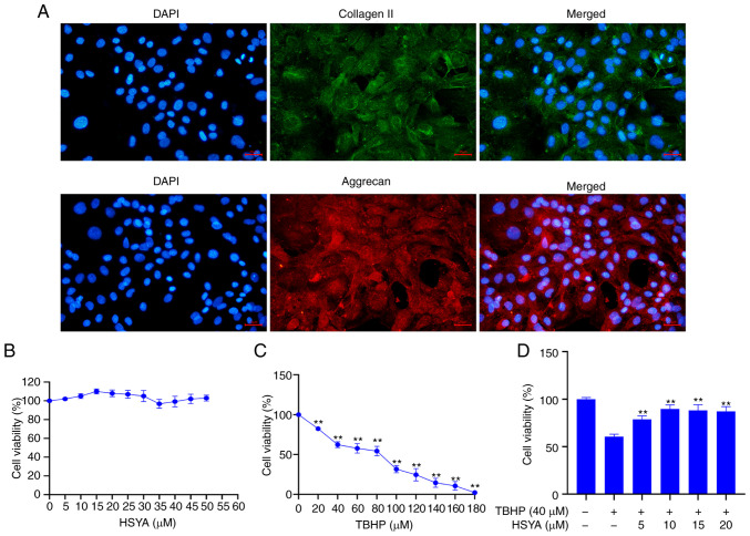
Figure 1.
Determination of drug concentration. (A) Immunofluorescence was used to confirm primary NP cells using collagen II and aggrecan. Scale bar, 25 µm. A CCK-8 assay was used to detect the effect of different concentrations of (B) HSYA and (C) TBHP on NP cell viability. **P<0.01 vs. 0 µM. (D) A CCK-8 assay was used to detect the effect of TBHP and HSYA on NP cell viability. **P<0.01 vs. 0 µM HSYA+TBHP. HSYA, hydroxysafflor yellow A; NP, nucleus pulposus; TBHP, tert-butyl hydroperoxide; CCK, Cell Counting Kit; DAPI, diamidino-2-phenylindole.

