Table 1.
Proposed limits of PTV fo r different osteoarticular locations
| SUPERIOR | INFERIOR | MEDIAL | LATERAL | PROXIMAL | DISTAL | |
|---|---|---|---|---|---|---|
|
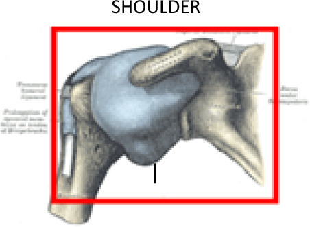
POST: 1 cm above acromion ANT: coracoid process |
1.5 cm under lesser tubercle | POST: 1.5 cm scapula ANT: 1.5 cm clavicle |
Lateral side-of humeral head, neck and epiphysis and 1 cm of surrounding soft tissues | ||
|
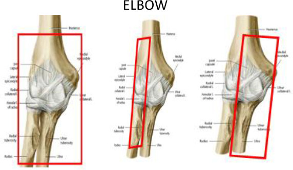
1.5 cm above trochlea and capitellum | 1.5 cm below radial and ulnar tuberosities | 1.5 cm around bone edges into surrounding soft tissues | 1.5 cm around bone edges into surrounding soft tissues | ||
|
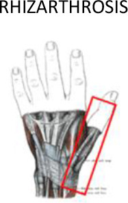
1 cm around of the soft tissues surrounding the bones | 1 cm around of the soft tissues surrounding the bones | Half of the metacarpal bone, the joint with the trapezoid bone and 1 cm through the radial bone | Proximal third of the distal phalanx | ||
|
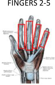
2 mm inside the dorsal skin | 2 mm inside the ventral skin | 1 cm around of the soft tissues surrounding | 1 cm around of the soft tissues surrounding |
|
|
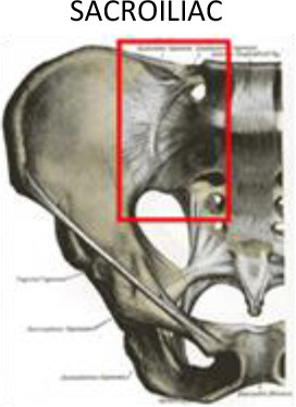
|
1 cm above sacroiliac joint | 1 cm below sacroiliac joint | 1 cm into sacral bone | 1 cm into iliac bone | ||
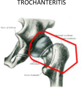
|
Trochanter, femoral head and neck | Below lesser trochanter | 1 cm around of the soft tissues surrounding | 1 cm around of the soft tissues surrounding | ||
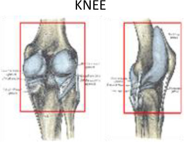
|
2 cm above femoral condyles | 2 cm below tibial condyles and fibula head | 1 cm around of the soft tissues surrounding | 1 cm around of the soft tissues surrounding | 1 cm above lateral and medial femoral epicondyle | Lateral and medial tibial condyles and fibula head |
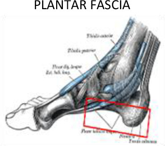
|
1 cm around of the soft tissues surrounding | 1 cm around of the soft tissues surrounding | Metacarpal-phalanx joint | Calcaneus tuberosity | ||
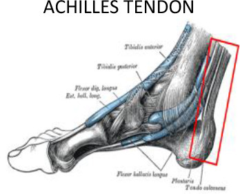
|
1 cm around the tendon | 1 cm around the tendon | Lateral and medial malleolus | Calcaneus tuberosity, subcutaneous calcaneus bursa, submuscular calcaneus bursa | ||
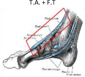
|
1 cm around the tendon | 1 cm around the tendon | 2 cm above medial malleolus |
|
||
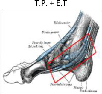
|
1 cm around the tendon | 1 cm around the tendon | 2 cm above lateral malleolus |
|
||
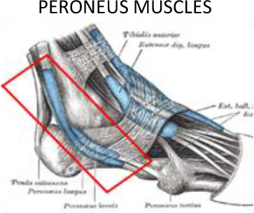
|
1 cm around the tendon | 1 cm around the tendon | 2 cm above lateral malleolus | fifth metatarsal bone, lateral side. |
E.T, Extensor tendons of the toes; F.T, Flexor tendons of the toes; T.A, Tibialis anterior; T.P, Tibilais posterior.
