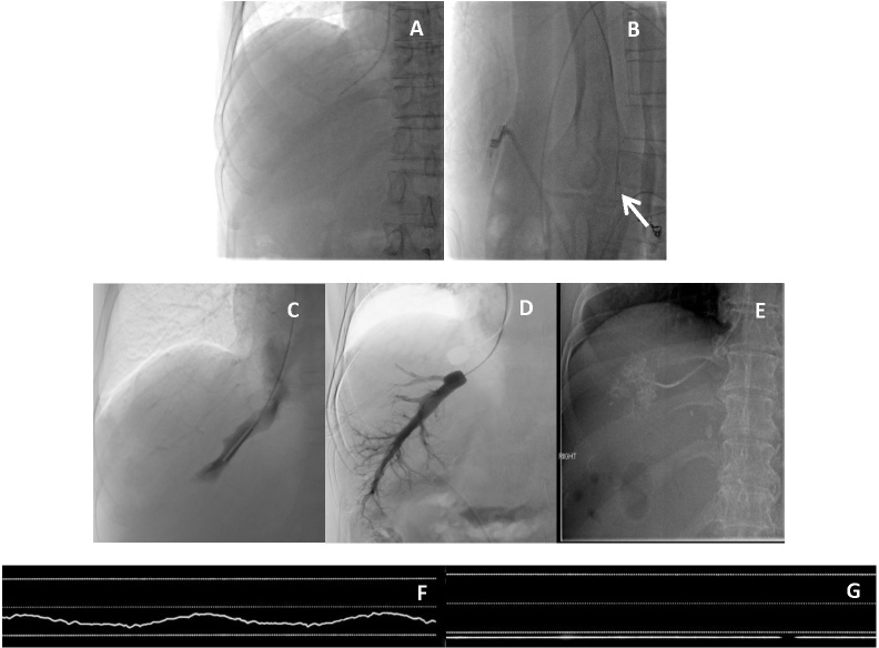
Figure 1.
Routine measurement of HVPG. Anterior view (a) and lateral view (b) showing the catheter coursing posteriorly in lateral view (arrow) indicating cannulation of the right hepatic vein. Measurement of FHVP and WHVP. (c): Balloon deflated to measure FHVP; d): Balloon inflated to measure WHVP, digital subtraction venogram confirmed good vessel occlusion and absence of HV–HV shunts. (e): Example of WHVP measurement by wedging catheter against a small hepatic vein branch in a different patient. (f) Sample waveform of FHVP measurement; g) Sample waveform of WHVP measurement. FHVP; free hepatic venous pressure; HVPG, hepatic venous pressure gradient; WHVP, wedged hepatic venous pressure.

