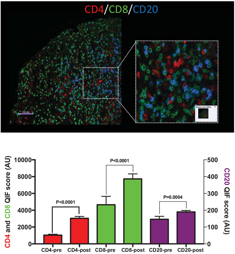
Figure 1.
Tumor infiltrating lymphocytes at baseline and after neoadjuvant radiotherapy in clinical sarcoma specimens. (A) An example of multiplexed immunofluorescence staining for tumor infiltrating CD8+ or CD4+ T cells and CD20+ B cells. (B) Quantification of tumor infiltrating lymphocyte staining before (pre) and after (post) neoadjuvant radiotherapy. Paired baseline biopsy preradiotherapy and tumor resection postneoadjuvant radiotherapy samples from 30 patients treated at UC Davis were used to generate a tissue microarray. Samples were stained with DAPI, anti-CD4, anti-CD8, anti-CD20, and anti-Cytokeratin at the Schalper laboratory.

