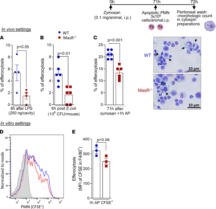
Figure 8. MasR is important for efferocytosis of apoptotic neutrophils.
WT and MasR–/– mice received an i.pl. injection of LPS (250 ng/cavity) (A) or were infected with E. coli intraperitoneally (B), and efferocytosis was morphologically identified in cytospin slides at 8 and 6 hours postchallenge, respectively. The frequency of efferocytosis was evaluated by counting 500 cells per slide. WT and MasR–/– were also used for the efferocytosis assay post-zymosan intraperitoneal injection, as shown in experimental design above the figure. Percentage of efferocytosis was obtained by morphological identification in cytospin slides (C). Representative images of the slides are shown in C. Original magnification, 100×. Arrows represent macrophages with engulfed apoptotic cells. Data are shown as the mean ± SEM of n = 4–6 mice in each group. Lastly, CFSE-labeled neutrophils were coincubated with WT or MasR–/– BMDMs for 1 hour, and flow cytometry was performed for efferocytosis evaluation (MFI of CFSE in macrophages — D and E). PMN, polymorphonuclear cell.

