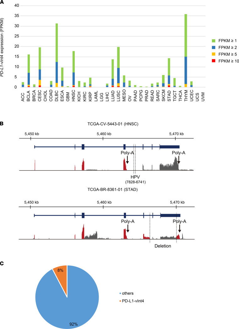
Figure 3. PD-L1–vInt4 expression was found in various types of cancer.
(A) The percentage of cases expressing PD-L1–vInt4–specific sequence in intron 4 with FPKM ≥ 1, ≥ 2, ≥ 5, and ≥ 10 in each TCGA cancer type. (B) Structural changes associated with PD-L1–vInt4 expression in a case of HNSCC (TCGA-CV-5443-01) and STAD (TCGA-BR-8361-01). Aberrant transcripts are colored in red. (C) PD-L1–vInt4 was detected in some of the clinical samples. Data were obtained from CCLE.

