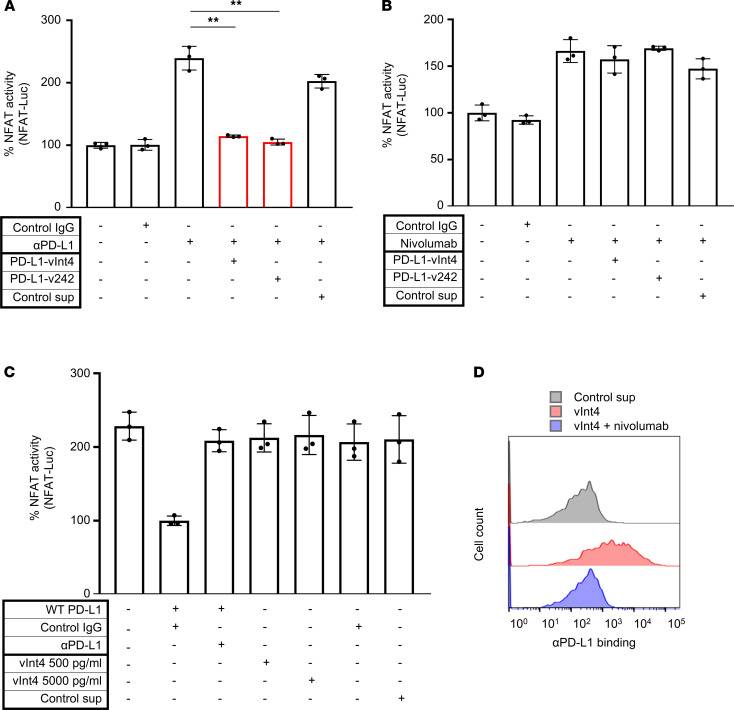
Figure 5. PD-L1–vInt4 demonstrated its function as a decoy in vitro.
(A) Evaluation of NFAT activity by measuring luminescence when PD-1 effector cells preincubated with αPD-L1 and secreting PD-L1 variants were cocultured with K1_PD-L1 WT cells. PD-L1–v242 and αPD-L1 were added in 3:1 molar ratio, whereas the ratio was 6:1 with PD-L1–vInt4 and αPD-L1. n = 3. **P < 0.01 by paired 2-tail Student’s t test. (B) Comparison of NFAT activity by measuring luminescence — this time using PD-1 effector cells preincubated with nivolumab and secreting PD-L1 variants. Secreted variants do not significantly suppress the signaling; paired 2-tail Student’s t test. Each condition was compared with the third bar counted from the left end in A and B. (C) Evaluation of NFAT activity by measuring luminescence. PD-1 effector cells were cocultured with K1 or K1_PD-L1 WT cells. PD-L1–vInt4 was added at a physiologically plausible concentration and at a 10-fold concentration. Secreted variants do not significantly suppress the signaling; paired 2-tail Student’s t test. Each condition was compared with the bar in the left end in C. Data represent mean ± SEM in A–C. (D) Jurkat–PD-1 cells incubated with or without Fc-tagged PD-L1–vInt4 were evaluated with FACS. As for nivolumab, it was added 30 minutes before PD-L1–vInt4.

