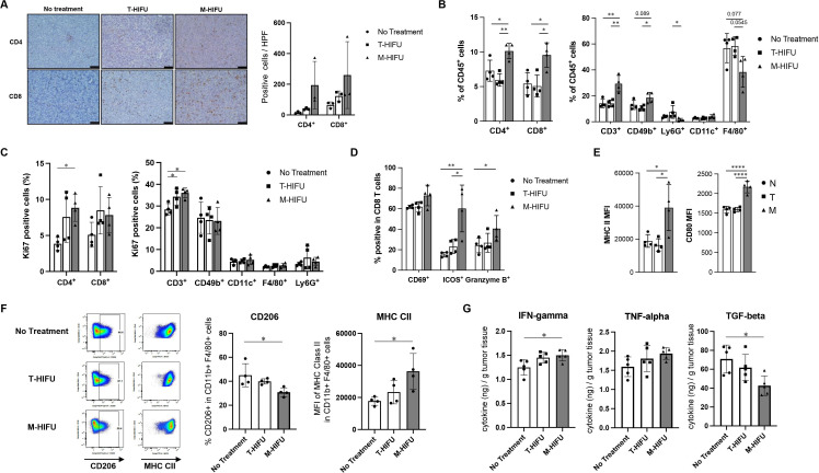
Figure 2.
Enhanced intratumoral infiltration of activated CD4+ and CD8+ cells and M1 polarized macrophages by M-HIFU. 1×10⁶ MM3MG-HER2 cells were injected into legs of BALB/c mice. Established leg tumors were treated with M-HIFU or T-HIFU on day 7 after tumor inoculation. (A) Immunohistochemical staining of T cells in tumor sections. Tumors on day 13 after HIFU treatment were collected, fixed with formalin and stained with anti-mouse CD4 and CD8 monoclonal antibodies. Representative images are shown (left: no treatment, middle: T-HIFU, right: M-HIFU). Scale bar is 50 μm. Quantification of positive cells in high-power field (HPF) is shown in the right panel. Error bars represent SD, n=3 per group. (B) Seven days after HIFU treatments of MM3MG-HER2 tumors in mice, tumors were collected and digested for flow cytometry analysis. The percentages of CD4+, CD8+, CD3+, CD49b+, Ly6G+, CD11c+, and F4/80+ cells in alive CD45+ cells were analyzed for each HIFU treatment group. n=4 per group. (C) The expression of proliferation marker Ki67 was analyzed for each cell type in tumor-infiltrating immune cells by flow cytometry, and percentages of Ki67 positive for each cell type are shown. n=4 per group. (D) The expression of CD69+, Inducible T-cell costimulator (ICOS)+, and granzyme B+ by CD8+ cells were analyzed by flow cytometry and shown for each HIFU treatment group. n=4 per group. (E) MFI of MHC class II (left) and CD80 (right) expression on CD11c+ dendritic cells are shown for each HIFU treatment group. (F) Expression of CD206 and MHC class II by CD11b+F4/80+ macrophage population was analyzed for each treatment group. Representative dot plots of CD206 and MHC class II staining are shown in the left panel. Percentages of CD206-positive macrophages and mean fluorescence intensity of MHC class II expression are shown. n=4 per group. (G) ELISAs for IFN-γ, TNF-α, and TGF-β1 were performed with tumor lysates made from MM3MG-HER2 tumors treated with no treatment, T-HIFU or M-HIFU. n=5 per group. Error bars represent SD. *P<0.05, **P<0.01, ****P<0.0001. IFN-γ, interferon gamma; MFI, mean fluorescent intensity; M-HIFU, mechanical high-intensity focused ultrasound; TGF-β1, transforming growth factor beta 1; T-HIFU, thermal high-intensity focused ultrasound; TNF-α, tumor necrosis factor alpha.

