. 2022 Jan 3;36(1):198–205. doi: 10.21873/invivo.12691

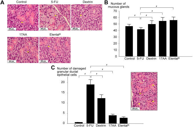
Figure 4. Histological changes of salivary glands in mice. (A) HE staining. Mucous gland size of the 5-FU group was bigger than that of the other groups. The yellow arrows show the mucous gland area of salivary gland. (B) Change of mucous gland number. The number of mucous glands was higher in the Control, Dextrin, 17AA and Elental® groups than that in the 5-FU group, whereas the 17AA and Elental® groups showed the highest number. #p<0.01; compared to the 5-FU group (Mann-Whitney U-test). (C) Change of damaged granular ductal epithelial cell number. The number of damaged granular ductal epithelial cells was higher in the 5-FU group than in all groups, and was lowest in the Elental® and 17AA groups. The yellow arrows show the disappearance or atrophy of granular ductal epithelial cells after 5-FU treatment. Bars, SD. #p<0.01; compared to the 5-FU group (Mann-Whitney U-test).

Figure 4