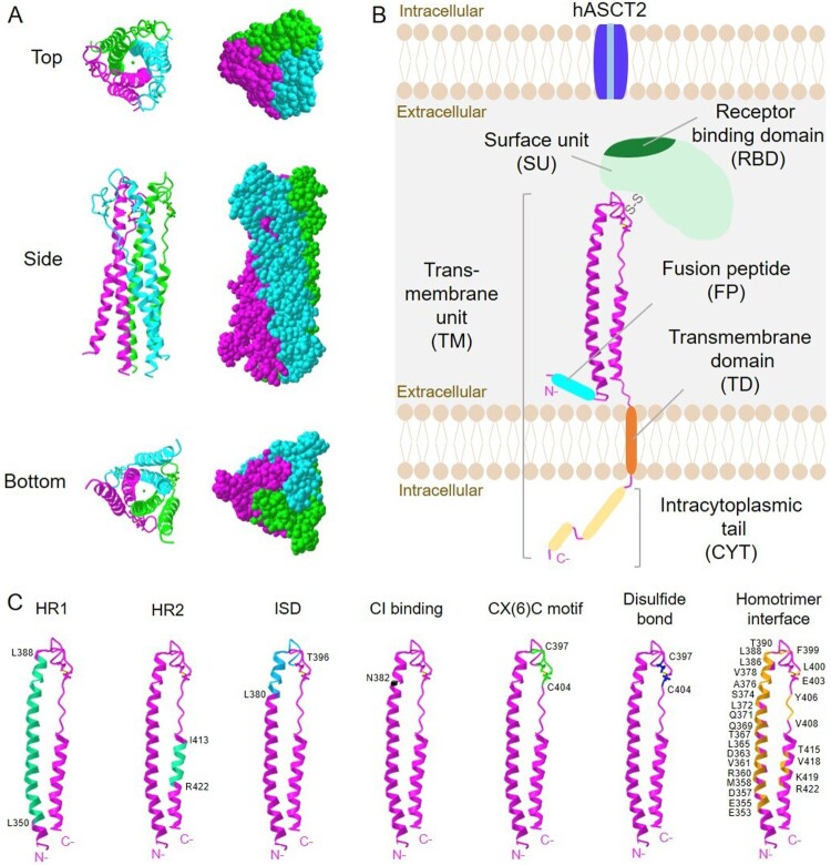
Figure 1.
Structure of syncytin-1. (A) 3D crystal structure images of trimeric syncytin-1 are created with iCn3D structure viewer (Wang et al. 2020). PDB ID: 5HA6 (doi:10.2210/pdb5HA6/pdb). (B) Schematic representation of syncytin-1 monomer describing the surface unit (SU) and transmembrane (TM) units. Crystalized region shown in (A) is described in pink ribbon. The receptor binding site (RBD) is located in SU, and binds to the hASCT2 receptor to trigger membrane fusion. One disulfide bond links SU and TM. Fusion peptide (cyan blue), transmembrane domain (orange), and the intracytoplasmic tail (yellow) are indicated. (C) Functional sites in syncytin-1 are highlighted. Amino acid residues forming heptad repeats (HR1 and HR2), immunosuppressive domain (ISD), CI binding site, CX(6)C motif, a disulfide bond and homotrimer interface are labeled.

