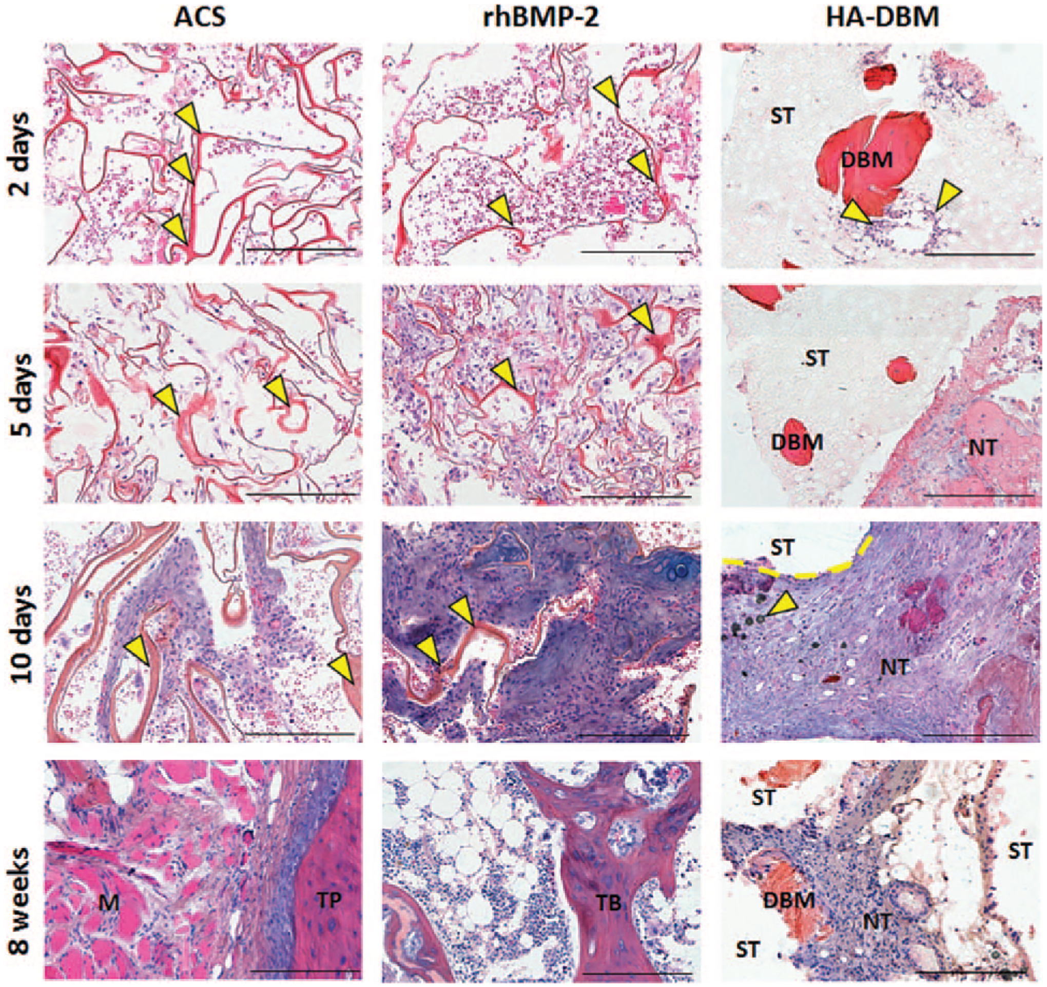
Figure 4.

Histological evaluation of the implants 2, 5, 10 days and 8 weeks postoperatively, stained with Gill’s hematoxylin, eosin and alcian blue. Yellow arrows in the ACS and rhBMP-2 samples indicate collagen bundles (red, ribbon-like). Yellow arrows in the 2 days HA-DBM sample indicate cells (purple nuclei) in proximity of a DBM particle. Yellow arrows in the 10 days HA-DBM sample indicate residual HA particles. Legend: Scaffold struts = ST; demineralized bone matrix = DBM; new tissue = NT; muscle M; transverse process = TP; trabecular bone = TB. Scale bars: 400 μm. ACS indicates absorbable collagen sponge; DBM, demineralized bone matrix; HA, hydroxyapatite.
