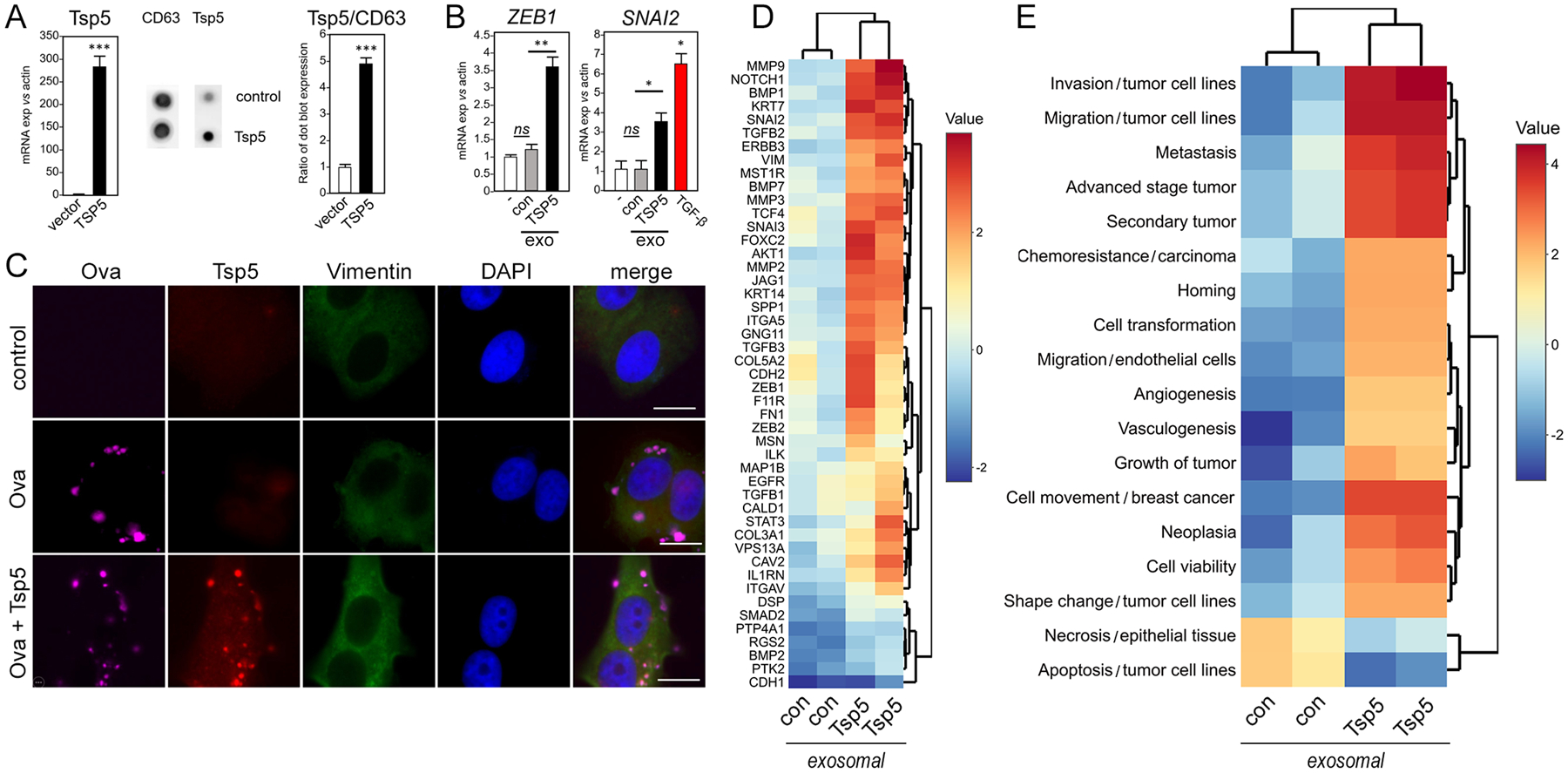
Fig. 4. TSP5 protein drives expression of EMT phenotypes.

Human pre-adipocytes were transduced with TSP5-expressing lentivirus or lentivirus vector control and differentiated to mature adipocytes as above. (A) RT-PCR of adipocyte total RNA was used to compare gene expression in TSP5-transduced adipocytes to vector control. Exosomes werepurified from conditioned media of transduced adipocytes and characterized by Elispot for expression of TSP5 protein and exosomal surface marker CD63. Exosomal TSP5 expression is reported as ratio with CD63. Data are from N=3 independent experiments; *** P <0.001 by unpaired, two-tailed t-test. (B) Upon addition to MCF7 cells, exosomes (exo) from adipocytes transduced with TSP5 lentivirus were compared to exosomes from adipocytes transduced with lentivirus vector control (con) for ability to induce transcription of EMT genes ZEB1 and SNAI2. Induction of SNAI2 by human recombinant transforming growth factor (TGF)-β is shown as a positive control for a pro-EMT response (red bar). Expression in each experimental was compared to vehicle control (−) using unpaired, two-tailed t-test. Data are from N=3 independent experiments. * P <0.05, ** P <0.01, and ns not significant by unpaired, two-tailed t-test. (C) MCF7 cells were treated for 3 days with synthetic cationic vesicles loaded with recombinant human TSP5, then vimentin expression was measured by immunofluorescence imaging as a measure of EMT induction. Anti-TSP5 antibody confirmed delivery of protein by the synthetic vesicles. As a control for loading and delivery, AlexaFluor 647-labeled ovalbumin (Ova) was also incorporated into the vesicles. For each of N=3 independent experiments, 25 images were examined by immunofluorescence. One representative image is shown, out of 25 images collected for each of the three experimental conditions with three independent replicates. Scale bar, 10 μm. (D) Heatmap of EMT gene expression by PCR array. MCF7 cells were treated for 5 days with exosomes purified from conditioned media of human primary adipocytes that had been transduced with either lentivirus overexpressing TSP5 or lentivirus vector control. Color scale bar shows expression range (data file S5). (E) IPA analysis of (D) generated Z scores of pathways and functions (data file S6).
