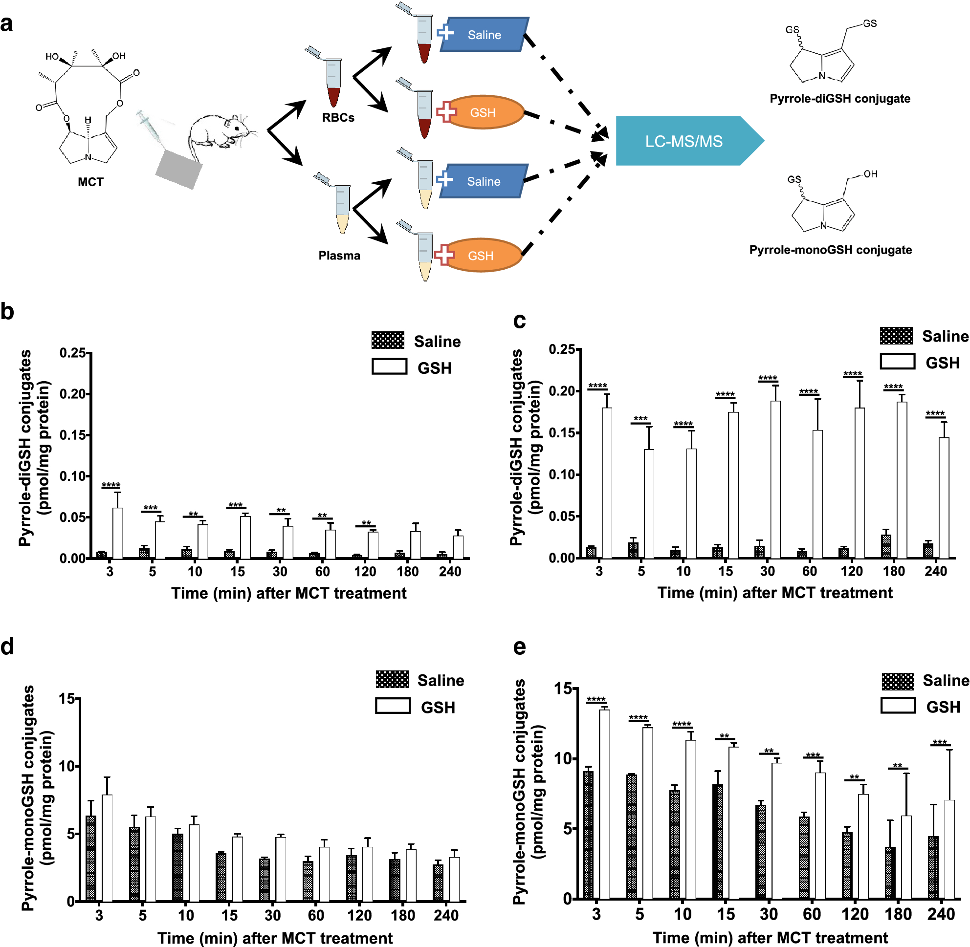
Fig. 6.

Detection of reactive metabolites of MCT in liver efflux blood of MCT-treated rats. Adult male SD rats (200–220 g) were anesthetized and treated with a single administration of MCT via intravenous injection at 65 mg/kg, and blood efflux from the liver (400 μL) was collected via the cannulated vein at various times (0–240 min) for further processing as illustrated (a). Pyrrole-diGSH conjugates (b, c) and pyrrole-monoGSH conjugates (d, e) were determined in RBCs (b, d) or plasma (c, e). Data are presented as means ± SD (n=3). Friedman test was used to compare GSH-added RBCs or plasma with saline-vehicle RBCs or plasma. **p<0.01, ***p<0.001, ****p<0.0001.
