FIG 9.
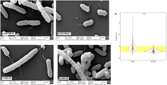
Electron microscopy of P. aeruginosa clone C bacteria. (A) SG17M, mid-exponential phase; (B) SG17M, early stationary phase; (C) NN2, mid-exponential phase; (D) NN2, early stationary phase. (E) Distribution of bacterial cell length of two biological replicates per strain and time point. ****, P < 0.0001.

