FIG 1.
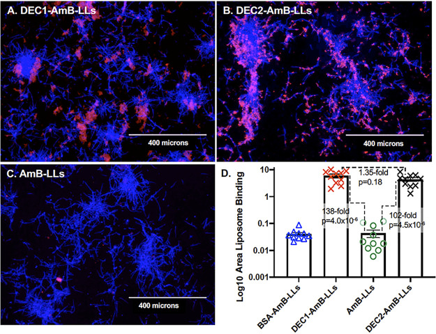
DEC1-AmB-LLs and DEC2-AmB-LLs bound specifically to in vitro grown C. albicans hyphae. C. albicans hyphae were stained with rhodamine tagged liposomes (A) DEC1-AmB-LLs and (B) DEC2-AmB-LLs, and (C) AmB-LLs. An image of BSA-AmB-LL binding is not shown. (A to C) Fungal cell chitin was stained with CW. The epifluorescence of chitin (blue) and liposomes (red) was photographed at ×10 magnification. All four preparations of liposomes were diluted equivalently. Size bars represent 400 microns. (D) A scatter bar plot compares the area of red fluorescent staining quantified from 10 images for each type of liposome. Standard errors and the fold differences in average area of staining and P values are indicated for comparisons of DectiSomes to AmB-LLs.

