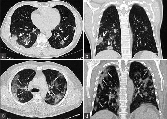
Figure 1.

(a and b) A 49-year-old male patient with COVID-19 presenting with fever, dyspnea, sore throat and a lymphocyte count within the normal range (neutrophil to lymphocyte ratio: 1.0, lymphocyte to C-reactive protein ratio: 2.8). A computed tomography scan obtained 4 days after the onset of symptoms showed bilateral peripheral multifocal patchy consolidations predominantly in the lower lobes with subtle air bronchogram (thick arrows). (c and d) 52-year-old man with positive reverse transcription polymerase chain reaction for severe acute respiratory syndrome coronavirus 2 manifesting with initial symptoms of fever, cough, dyspnea, myalgia and lymphocytopenia (neutrophil-to-lymphocyte ratio: 11.1, lymphocyte-to-C-reactive protein ratio: 0.23). A computed tomography scan obtained 4 days after the onset of symptoms showed bilateral diffused peripheral ground glass opacities with crazy-paving pattern (thin arrows)
